MCQs on ECG

Written by Dr. Daniel Fraser (MBBS, FRACP) – Physician

Question 1

A 56-year-old man presents to the emergency department with complaints of chest pain and lightheadedness. On examination he has a blood pressure of 85/50 mmHg, rapid and barely perceptible pulse of 160 bpm and respiratory rate of 24 breaths per minute. An ECG is obtained which is shown in the accompanying photograph. Which one of the following is the most appropriate next step in management?
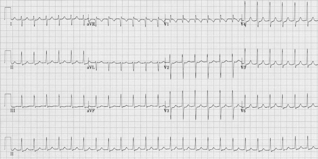
A) Atropine
B) Intravenous diltiazem
C) Intravenous metoprolol
D) Pace maker insertion
E) DC cardioversion

Correct Answer: E) DC cardioversion

The ECG is characteristic of supraventricular tachycardia (SVT)—tachycardia with regular and monomorphic QRS complexes and absence of P waves.

SVT can occur in individuals of all age groups with a spectrum of presentations ranging from asymptomatic to severely symptomatic. Symptoms may include palpitations, dizziness, shortness of breath, syncope, chest pain, fatigue, diaphoresis, and nausea.

Rare complications of paroxysmal SVT include myocardial infarction, congestive heart failure, syncope, and sudden death.

Acute management focuses on controlling the rate and preventing hemodynamic collapse. In hemodynamically unstable patients (hypotension, chest pain, dyspnea, altered perfusion), immediate DC cardioversion is the treatment of choice.

This patient has a systolic BP <90 mmHg, indicating hemodynamic instability, necessitating urgent synchronized cardioversion after appropriate sedation.

If the patient were stable, initial steps would include vagal maneuvers and adenosine. If those fail, AV nodal blocking agents like diltiazem or metoprolol may be used, targeting AVNRT or AVRT.

Options A and D (Atropine and pacemaker) are for symptomatic bradycardia, which is not relevant in this case.

Question 2

A 72-year-old woman presents to the clinic with complaints of intermittent palpitations and mild shortness of breath over the past month. She denies chest pain or syncope. On examination, her pulse is irregularly irregular with a rate of about 110 bpm. Blood pressure is 130/80 mmHg. An ECG is obtained as is as follows;

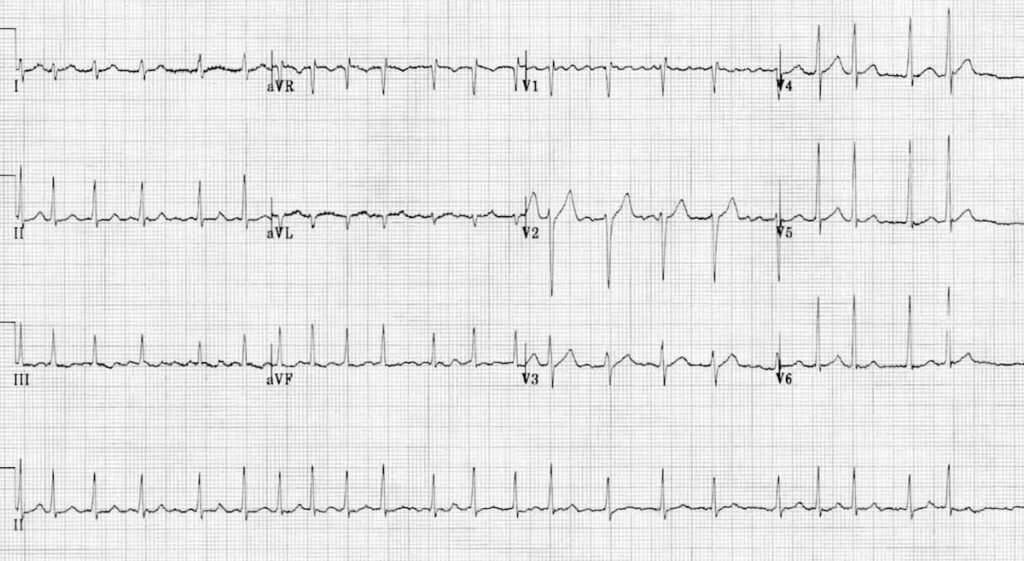
Which one of the following is the most likely diagnosis?

A) Atrial flutter
B) Atrial fibrillation
C) Sinus tachycardia
D) Ventricular tachycardia
E) Supraventricular tachycardia

Correct Answer: B) Atrial fibrillation

The ECG demonstrates several hallmark features of atrial fibrillation. There is an irregularly irregular ventricular rhythm, with variable R–R intervals and no repeating pattern. The baseline shows absence of discrete P waves, replaced instead by fine fibrillatory waves. This indicates that the atria are not contracting in a coordinated manner but are fibrillating chaotically.

Clinically, the patient has an irregularly irregular pulse and a ventricular rate around 110 bpm, which aligns with atrial fibrillation with a rapid ventricular response. Her symptoms of intermittent palpitations and mild shortness of breath are also classical for AF. The normal blood pressure and absence of chest pain or syncope make other arrhythmias less likely.

  • Atrial flutter (A) typically shows a “saw-tooth” pattern of flutter waves, usually with a regular ventricular response unless variable block is present.
  • Sinus tachycardia (C) would have a regular rhythm with visible P waves before each QRS.
  • Ventricular tachycardia (D) presents as a wide-complex tachycardia and is usually regular.
  • Supraventricular tachycardia (E) generally shows a regular, fast narrow-complex rhythm.

Because the strip clearly shows irregularly irregular narrow-complex tachycardia with absent P waves, atrial fibrillation is the most accurate diagnosis.

Question 3

A 60-year-old man presents to the Emergency Department with palpitations for the past 2 weeks. On examination, his blood pressure is 125/95 mmHg and his heart rate is irregular at 160 bpm. An ECG shows atrial fibrillation (AF). He is started on metoprolol for rate control, and electrical cardioversion is planned. Which one of the following should be used for anticoagulation?

A) Heparin, 24 hours before the procedure
B) Warfarin 4 weeks before, to 4 weeks after the procedure
C) Aspirin
D) Apixaban from 48 hours before, to 48 hours after the procedure
E) Flecainide

Correct Answer: B) Warfarin 4 weeks before, to 4 weeks after the procedure

Converting atrial fibrillation to sinus rhythm carries a risk of thromboembolism, primarily due to dislodgement of pre-existing left atrial thrombi formed during atrial blood stasis, or sometimes de novo thrombus formation around the time of cardioversion. This risk is particularly high in patients with AF lasting more than 48 hours, with an embolic risk of 1-5% in the first month if anticoagulation is not given.

To minimize this risk, prophylactic anticoagulation is recommended starting 4 weeks before and continuing 4 weeks after the cardioversion procedure. Oral anticoagulation with warfarin targeting an INR of 2-3 is the standard approach.

Heparin may be used initially with warfarin to prevent a prothrombotic state until the INR reaches the therapeutic range.

(Option A) Heparin alone is used in emergency cardioversion situations where immediate anticoagulation is needed, such as in hemodynamically unstable patients.

(Option C) Aspirin is inadequate for thromboembolism prevention in this context.

(Option D) While direct oral anticoagulants (DOACs) like apixaban and rivaroxaban have shown efficacy comparable or superior to warfarin, their safety and effectiveness specifically before and immediately after cardioversion are not fully established.

(Option E) Flecainide is an antiarrhythmic agent for pharmacological cardioversion and has no anticoagulant properties.

2. Cardiology Module