7.3 Skin Tumors

Question 1

John, 60 years of age, presents to your office because he is concerned about a skin lesion on his forearm. The dermatoscopic view of the lesion is shown in the following photograph. Which one of the following could be the most likely diagnosis?

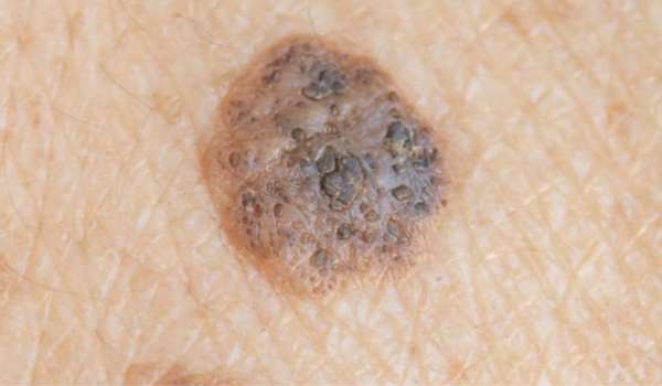
A) Actinic keratosis


B) Bowen’s disease
C) Malignant melanoma
D) Dermatosis papulosa nigra
E) Seborrheic keratosis

Correct Answer: E) Seborrheic keratosis

The photograph shows an oval, brown plaque on a sun-exposed area in an older adult. The lesion demonstrates keratotic plugging on the surface, appearing yellowish—a hallmark feature of seborrheic keratosis (SK).

Seborrheic keratosis is the most common benign skin tumor in older individuals. These lesions are typically oval or round, brown, and appear stuck on, often with a velvety or verrucous (warty) surface and adherent greasy scale. The color can range from tan to black.

  • Option A (Actinic keratosis): Usually presents as flat, scaly lesions on sun-exposed areas like the backs of the hands. It is a precancerous lesion that can progress to squamous cell carcinoma. Lesions are often multiple and less elevated than SK.
  • Option B (Bowen’s disease): Also occurs on sun-damaged skin in older adults and is precancerous, but the lesion is larger, thicker, flat, scaly, and has well-demarcated borders. It lacks the greasy, stuck-on appearance of SK.
  • Option C (Malignant melanoma): Typically presents as a flat lesion with multiple colors, irregular borders, and asymmetric shape. Though nodular melanoma can be elevated, it lacks keratotic plugs and often shows color variation, which is not seen in this case.
  • Option D (Dermatosis papulosa nigra): A variant of SK, but occurs in young to middle-aged individuals with darker skin tones, often presenting as multiple small papules on the face. This does not fit the clinical setting of a single lesion on the forearm of an older white male.

Thus, based on clinical appearance, location, age, and dermatoscopic features, the most likely diagnosis is seborrheic keratosis.

Question 2

A 45-year-old woman presents with a mole on the lower eyelid of her right eye as shown in the following photograph. Which one of the following would be the most appropriate management?

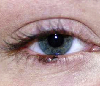
A) Excision of the lesion under local anesthesia.
B) Review in one month.
C) Reassure that the lesion is benign.
D) Refer the patient to a plastic surgeon.
E) Apply topical imiquimod.

Correct Answer: D) Refer the patient to a plastic surgeon.

The photograph shows an irregular pigmented lesion on the lower eyelid, raising suspicion for melanoma. In such cases, complete excision and histopathological examination are essential for diagnosis and staging.

Due to the anatomically delicate location, excision should be done by a plastic surgeon to minimize the risk of tissue destruction or functional impairment, particularly involving eyelid function and appearance.

  • (Option A) Excision under local anesthesia may be appropriate in general, but given the location, it should be performed by a specialist to ensure precision and optimal cosmetic/functional outcomes.
  • (Option B) Reviewing in one month delays definitive diagnosis and treatment, which is not acceptable in suspected melanoma.
  • (Option C) Reassurance is inappropriate as the lesion has malignant potential.
  • (Option E) Topical imiquimod is not suitable for suspected melanoma and is not recommended for use on eyelid skin.

Question 3

A 61-year-old male farmer presents with a lesion on his face. The lesion has a pearly, shiny surface with visible telangiectasia and some ulceration.

Which one of the following is the most likely diagnosis?

A) Keratoacanthoma
B) Basal cell carcinoma
C) Implantation dermoid cyst
D) Amelanotic malignant melanoma
E) Squamous cell carcinoma

Correct answer: B) Basal cell carcinoma.

Explanation:

  • The lesion’s pearly appearance, shiny surface, and visible telangiectasia are classic signs of nodular basal cell carcinoma (BCC), the most common skin cancer.
  • Ulceration in parts of the lesion is also common in BCC.
  • Keratoacanthoma typically has a central crater filled with necrotic tissue, absent here.
  • Implantation dermoid cysts are firm cystic nodules usually at sites of penetrating trauma like fingertips, not facial lesions.
  • Amelanotic melanoma can mimic nodular BCC but lacks the pearly appearance seen here.
  • Squamous cell carcinoma usually presents as an ulcerating plaque, which does not match this lesion’s features.

Author – Dr. James Whitfield (MBBS, FRACGP)

With over 30 years in primary care, Dr. James Whitfield is a highly experienced GP providing comprehensive medical services for individuals and families. He has a strong background in chronic disease management, preventive health, and minor surgical procedures.

7. Dermatology Module