12.3 Lower Limb Fractures

Question 1

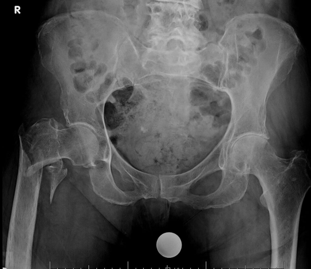
A 72-year-old woman presents to the emergency department after a fall at home. She complains of severe pain in her right hip and is unable to bear weight on the affected leg. On examination, the right lower limb is shortened, externally rotated, and there is tenderness over the hip region. An X-ray of the pelvis and right hip (see image) is obtained.

Which of the following best describes the diagnosis?

A) Intertrochanteric fracture of the femur
B) Femoral neck fracture
C) Acetabular fracture
D) Pubic ramus fracture
E) Hip dislocation

Answer: B) Femoral neck fracture


Explanation:

History and Clinical Examination:
Patients with a femoral neck fracture usually present with sudden onset hip pain following trauma, commonly a fall. The affected leg is often shortened and held in external rotation due to muscle spasm. Localized tenderness is present over the hip joint.

X-ray Features:
The diagnosis is confirmed on X-ray, which shows a fracture line through the femoral neck. Displacement can be seen as shortening and external rotation of the femur. This distinguishes it from other types of hip fractures, such as intertrochanteric fractures, which occur distal to the neck.

Management:
Management depends on the patient’s age, fracture displacement, and overall health. In elderly patients, displaced femoral neck fractures typically require surgical intervention, usually either hemiarthroplasty or total hip arthroplasty. Nondisplaced fractures may be managed with internal fixation using screws or pins. Early surgery is preferred to reduce complications such as avascular necrosis and nonunion. Postoperative rehabilitation with early mobilization is crucial for recovery.

Question 2

A 78-year-old woman presents after a fall at home with severe pain in her left hip and inability to bear weight. On examination, the left leg is shortened and externally rotated. Pelvic X-ray shows a displaced fracture through the femoral neck.

Which of the following is the most serious complication associated with femoral neck fractures?

A) Deep vein thrombosis
B) Avascular necrosis of the femoral head
C) Infection
D) Nonunion due to poor blood supply to the femoral shaft
E) Fat embolism syndrome

Correct Answer: B) Avascular necrosis of the femoral head

Femoral neck fractures disrupt the blood supply to the femoral head, mainly via the medial femoral circumflex artery. This can lead to avascular necrosis (AVN), a serious complication causing bone death and subsequent joint collapse. The risk of AVN is higher in displaced fractures and in younger patients due to disruption of the retinacular vessels.

Deep vein thrombosis (A) and infection (C) are possible postoperative complications but are less specific. Nonunion (D) is also a risk but AVN is more characteristic of femoral neck fractures. Fat embolism syndrome (E) is more common with long bone shaft fractures.

Early recognition and appropriate surgical management (hemiarthroplasty or internal fixation depending on patient factors) are critical.

Question 3

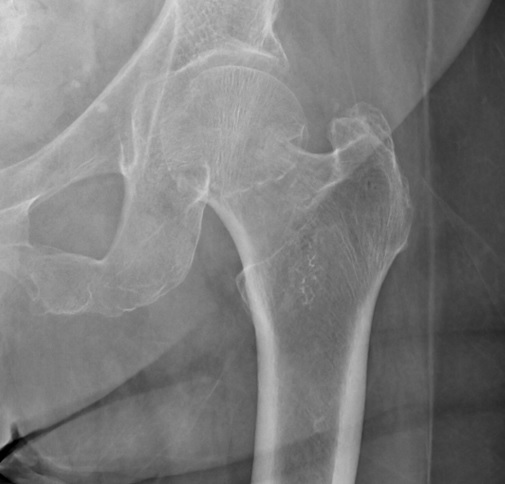
An 80-year-old woman slips on a wet floor and falls onto her right side. She presents to the emergency department with severe pain in her right hip and is unable to walk. On examination, her right leg appears shortened and externally rotated. There is tenderness and swelling around the greater trochanter. An X-ray of the right hip (see image) is performed.

Which of the following is the most likely diagnosis?

A) Femoral neck fracture
B) Intertrochanteric fracture
C) Subtrochanteric fracture
D) Acetabular fracture
E) Hip dislocation

Answer: B) Intertrochanteric fracture (right side)


Explanation:

History and Clinical Examination:
Intertrochanteric fractures typically occur in elderly patients following a low-energy fall, often onto the lateral hip. Patients present with acute hip pain, inability to bear weight, and swelling around the proximal femur. On physical exam, the affected limb is often shortened and externally rotated, similar to femoral neck fractures. Tenderness is maximal over the greater trochanter area, and swelling may be present due to soft tissue injury.

X-ray Features:
On the anteroposterior X-ray of the hip, intertrochanteric fractures are identified as fractures occurring between the greater and lesser trochanters of the femur. The fracture line usually runs obliquely or transversely through this region. These fractures are extracapsular, which influences blood supply and management decisions, and usually show displacement of the fragments.

Management:
Intertrochanteric fractures almost always require surgical fixation, commonly with a dynamic hip screw or an intramedullary nail. Early surgery followed by mobilization reduces complications such as pneumonia, deep vein thrombosis, and pressure sores. Conservative management is rarely indicated except in non-ambulatory or medically unfit patients.

12. Orthopedics Module