AMC Wise will be offline from 12.00am, 7/2/2026 to 8.00am 9/2/2026 for a scheduled maintenance.
Question 1
A 25-year-old man presents to the emergency department after falling off his bicycle onto his left shoulder. He reports immediate pain over the upper chest and difficulty moving his left arm. On examination, there is swelling and tenderness over the midshaft of the left clavicle, with visible deformity. Shoulder movement is limited due to pain, but distal neurovascular function is intact. An X-ray of the left clavicle is given below.
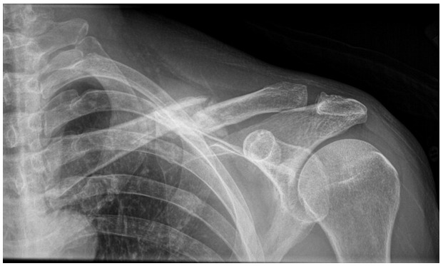
Which of the following is the most likely diagnosis?
A. Anterior dislocation of the shoulder
B. Clavicle fracture
C. Acromioclavicular joint separation
D. Proximal humeral fracture
E. Scapular fracture
Question 2
A 25-year-old man falls onto his outstretched hand during a rugby match and complains of pain and deformity over his right shoulder. On examination, there is tenderness over the middle third of the clavicle with a visible bump and limited shoulder movement. Neurovascular examination is normal.
Which of the following statements regarding clavicle fractures is correct?
A) The middle third is the most common site of fracture due to its relative anatomical weakness
B) Clavicle fractures frequently cause injury to the brachial plexus and subclavian vessels
C) Surgical fixation is routinely recommended for all clavicle fractures to prevent malunion
D) Clavicle fractures rarely heal without surgical intervention
E) Fractures of the lateral third are most commonly associated with pneumothorax
Question 3
A 72-year-old woman presents to the emergency department after tripping on a rug and falling onto her outstretched right hand. She reports severe pain in the right shoulder and upper arm. On examination, there is swelling, bruising, and marked tenderness over the proximal humerus. Passive and active shoulder movements are limited due to pain. Distal neurovascular examination is normal.
An X-ray of the right shoulder is given below.
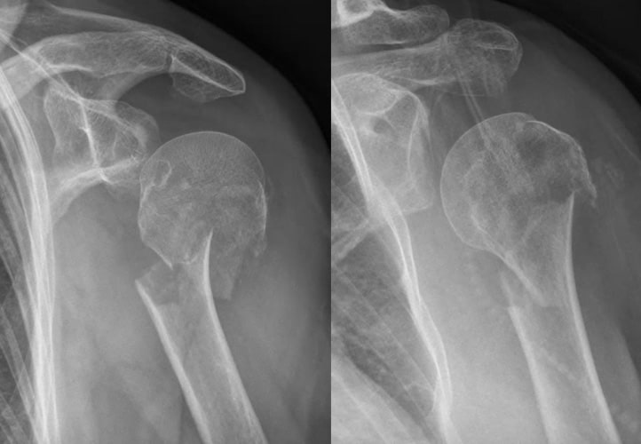
Which of the following is the most likely diagnosis?
A. Anterior shoulder dislocation
B. Clavicle fracture
C. Proximal humerus fracture
D. Rotator cuff tear
E. Acromioclavicular joint separation